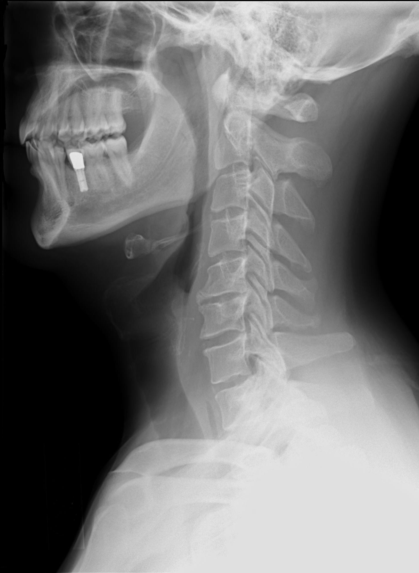
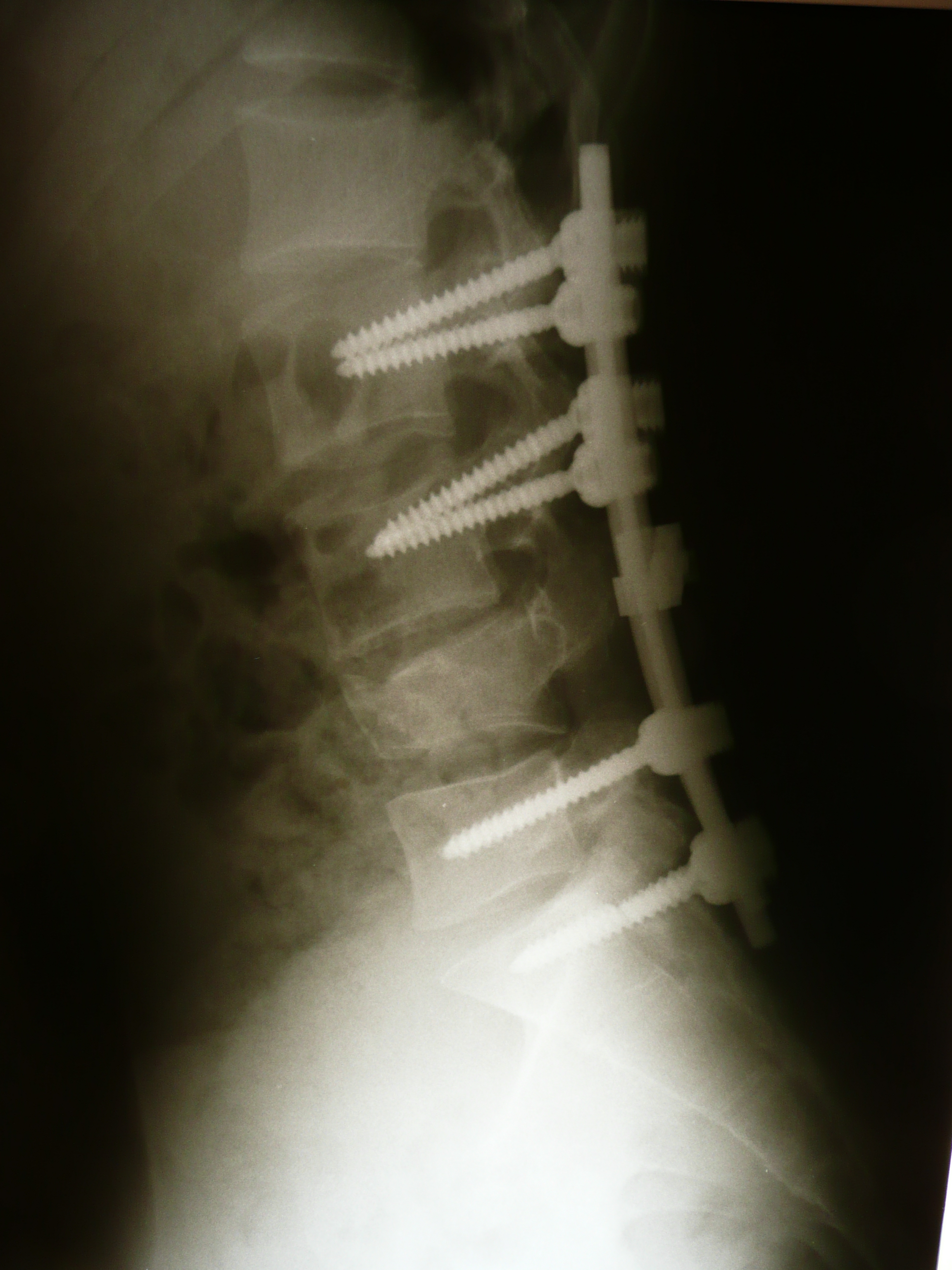
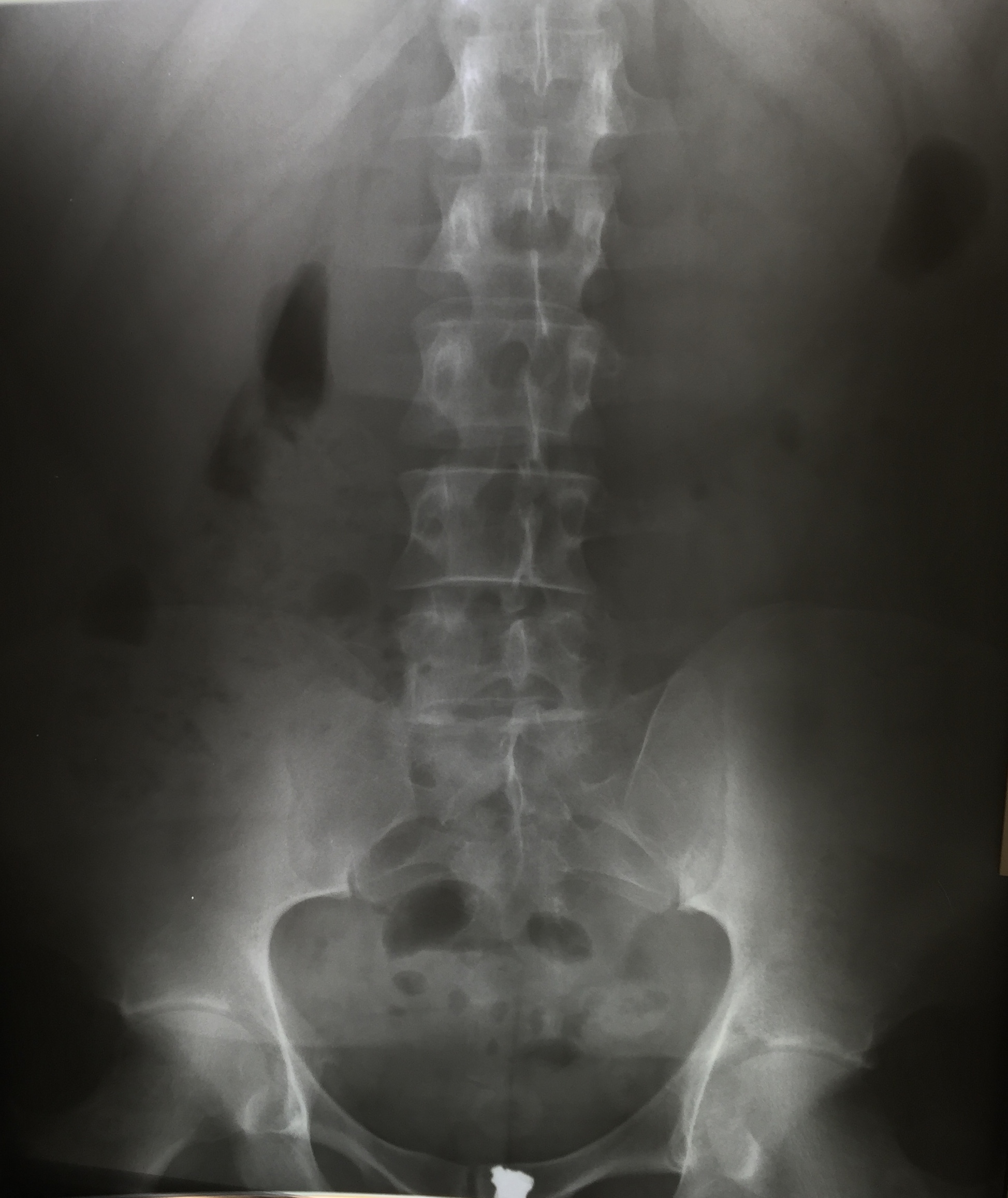
Educational images

This page provides open-license educational visuals for spine X-ray interpretation, minimally invasive procedures, and follow-up recovery discussion.

These materials are for education only and do not replace individualized diagnosis, treatment planning, or a physician consultation.

AP lumbar X-ray (example)

Spine X-ray

AP lumbar X-ray (example)

Frontal lumbar view used to review vertebral alignment and bony changes.

Clinical relevance: Useful for screening alignment issues; often interpreted with symptoms and exam findings.

License: CC BY-SA 4.0 View source →
Lateral lumbar X-ray (example)

Spine X-ray

Lateral lumbar X-ray (example)

Side-view lumbar radiograph for disc space height, sagittal profile, and degenerative changes.

Clinical relevance: Helps clinicians understand mechanical stress patterns in the lumbar spine.

License: CC BY-SA 4.0 View source →
Cervical lateral X-ray (pre-surgical evaluation)

Spine X-ray

Cervical lateral X-ray (pre-surgical evaluation)

Lateral cervical radiograph used in preoperative assessment for instability and degeneration.

Clinical relevance: Dynamic or multi-view cervical studies can guide whether surgery is appropriate.

License: CC0 (public domain dedication) View source →
Lumbar laminectomy illustration

Minimally invasive procedure

Lumbar laminectomy illustration

Educational diagram explaining decompression of neural structures in the lumbar region.

Clinical relevance: Supports discussion of goals: nerve decompression, pain relief, and functional recovery.

License: CC BY-SA 4.0 View source →
ACDF coronal educational diagram

Minimally invasive procedure

ACDF coronal educational diagram

Annotated coronal-style diagram for understanding cervical decompression and fusion concepts.

Clinical relevance: Helpful for explaining anatomy, compression pattern, and why fusion may be recommended.

License: CC BY-SA 3.0 / GFDL View source →
Follow-up radiology view (instrumentation example)

Recovery and follow-up

Follow-up radiology view (instrumentation example)

Post-treatment style radiology image showing hardware and alignment for follow-up interpretation.

Clinical relevance: Follow-up films are used with symptom tracking and physical function milestones.

License: CC BY-SA 3.0 (or compatible listed license) View source →

How to use these images in patient education

  • Start with plain-language anatomy before discussing pathology.
  • Pair each image with symptoms, neuro exam, and functional impact.
  • Explain expected goals of conservative and surgical options.
  • Use follow-up imaging alongside pain and mobility outcomes.
Ask your care team a question